Abdomen and Pelvis Sonography: Everything You Need to Know
So your doctor just handed you a referral slip that says “USG abdomen and pelvis.” And now you’re staring at it wondering, what exactly happens during this test? Will it hurt? Do I have to starve? What do the results even mean?
Relax. You’re not alone, and this guide has you covered.
Whether you’re searching for a reliable 5D Ultrasound machine test in Delhi or just trying to understand what abdomen and pelvis sonography is all about, read on. We’ll walk through everything in plain English.
What Is Abdomen and Pelvis Sonography?
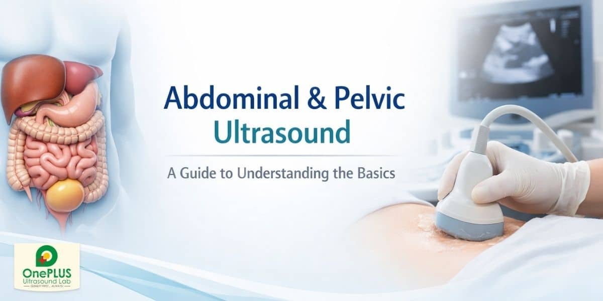
Abdomen and pelvis sonography (also called USG abdomen and pelvis) is a non-invasive imaging procedure that uses high-frequency sound waves to create real-time images of your internal organs.
No radiation. No needles. No drama.
The scan examines organs like the liver, kidneys, gallbladder, spleen, pancreas, bladder, and reproductive organs. Doctors use it to detect conditions ranging from kidney stones to tumours to cysts, and a whole lot more.
According to the American Institute of Ultrasound in Medicine (AIUM), abdominal ultrasound is one of the most widely used diagnostic tools globally because of its safety, accuracy, and accessibility.
Why Does Your Doctor Order This Scan?
There’s usually a good reason, and it’s rarely the worst-case scenario your brain jumps to at 2 AM.
Common reasons your doctor may refer you for USG abdomen pelvis include:
- Persistent abdominal pain or bloating
- Suspected gallstones or kidney stones
- Abnormal liver function test results
- Evaluation of pelvic pain in women
- Monitoring cysts, fibroids, or ovarian issues
- Urinary tract problems or enlarged prostate in men
- Follow-up after previous findings
The ultrasound imaging of abdomen and pelvis gives your doctor a real-time map of what’s happening inside, without any surgical intervention. That’s genuinely remarkable when you think about it.
How to Prepare for Abdomen and Pelvis Sonography
This is probably the most common question people search for, and honestly, preparation can make or break the quality of your scan.
Here’s what you need to know:
For abdominal organs ultrasound scan:
- Fast for at least 6 to 8 hours before the test. Food and drinks (especially fatty ones) cause the gallbladder to contract, making it harder to visualise clearly.
- You can take your regular medications with a small sip of water.
- Avoid carbonated drinks and chewing gum the night before, gas makes your sonographer’s job much harder.
For pelvic organs sonography:
- Drink 2 to 3 glasses of water about 1 hour before the test and don’t urinate.
- A full bladder acts as an acoustic window that helps the sound waves travel better, giving clearer images of the uterus, ovaries, and bladder.
Pro tip: Wear comfortable, loose clothing. You’ll be asked to expose your abdomen and lower pelvic region, and a hospital gown is often provided.
What to Expect During Ultrasound Abdomen and Pelvis Scan
Once you arrive at the diagnostic centre, the process is straightforward and typically takes about 20 to 45 minutes.
Here’s the step-by-step:
- You lie on an examination table, usually on your back.
- The sonographer applies a clear, water-based gel to your abdomen. It feels slightly cold, this is the only “discomfort” most people report.
- A handheld device called a transducer is pressed gently against your skin and moved in different directions.
- Sound waves bounce off internal structures and return as electrical signals that form images on a monitor.
- The sonographer captures still images and short video clips for the radiologist to review.
- Once done, the gel is wiped off. You’re free to eat, drink, and continue your day normally.
That’s it. No needles. No recovery time. No side effects.
At OnePLUS Ultrasound Lab, we use advanced ultrasound machines, including 5D Ultrasound machine test in Delhi, to ensure high image resolution and diagnostic accuracy. You can find our location easily on Google My Business by searching OnePLUS Ultrasound Lab.
Understanding USG Abdomen Pelvis Results Step by Step
Getting your ultrasound report can feel like reading a foreign language. Here’s a quick breakdown:
Normal findings will often describe organs as “normal in size, shape, and echotexture”, which is a good thing.
Echogenicity refers to how bright or dark a structure appears on the scan. Abnormal echogenicity in the liver, for instance, can suggest fatty liver disease.
Common findings you might see in the report:
- Cholelithiasis: gallstones
- Renal calculi: kidney stones
- Hepatomegaly: enlarged liver
- Cystic lesion: fluid-filled sac (often benign)
- Free fluid in pelvis: may indicate infection, injury, or other conditions
Always read the report with your doctor, not instead of them. A radiologist describes; your physician diagnoses. These are two different steps in the process.
Benefits of Abdominal and Pelvic Ultrasound for Diagnosis
Why is abdomen sonography still the go-to test despite MRI and CT scans being available?
Because it’s:
Safe: No ionising radiation means it is a choice of pregnant women, children and patients who require frequent monitoring.
Fast: This is a complete abdominal and pelvic ultrasound that can usually be completed in 30-45 minutes.
Affordable: USG abdomen scanning is much cheaper than MRI and CT and does not compromise detection of most conditions.
Live imaging: The physicians can see movement of organs, blood flow with Doppler mode, and foetal heartbeat in real-time, not on some static images.
Widely available: Quality ultrasound units can be found in even smaller cities and clinics, which is not the case with advanced scanners.
In the case of the North Delhi population, OnePLUS Ultrasound Lab, a reputable Diagnostic Centre in North Delhi, provides a good quality of abdominal and pelvic ultrasound services, with qualified radiologists present. Complementing the imaging unit are our Pathology Lab in Pitampura, and you can get blood tests and scans in a single building without having to run between them.
Who Should Get an Abdomen and Pelvis Sonography?
In short, anyone their doctor recommends it for. But here are specific groups who commonly need it:
- Women with irregular periods, pelvic pain, suspected PCOS, endometriosis, or early pregnancy evaluation
- Men over 40 for prostate and bladder assessment
- People with diabetes or high blood pressure for kidney and liver monitoring
- Anyone with digestive complaints lasting more than a few weeks
- Patients on long-term medications that affect the liver
Annual wellness scans are also increasingly common, many people now include abdomen sonography in their regular health check-up package. Prevention is genuinely cheaper than cure.
How Often Should You Get This Test?
There’s no universal rule. Your doctor will guide you based on your symptoms and health history.
For healthy adults with no symptoms, a routine abdominal scan every 2–3 years after age 40 is reasonable, especially if you have a family history of liver disease, kidney issues, or gallbladder problems.
If you’re already being treated for a condition, your doctor may recommend follow-up scans every 3 to 6 months to track changes.
Conclusion: Key Takeaways on Abdomen and Pelvis Sonography
The sonography of the abdomen and pelvis is not to be feared, but used. It is one of the strongest, most convenient, and safe instruments of the new diagnostics that allow physicians to check your inner organs without a single cut.
You need to comprehend a referral, know how to prepare abdomen and pelvis sonography, or get your report straight, this guide provides you with a straightforward and realistic point of departure. And when you are familiar with what to anticipate when you have to do an ultrasound scan on the abdomen and pelvis, the entire process becomes a lot easier.👉 Don’t delay your health check. Book your ultrasound abdomen and pelvis today and get the clarity you deserve, because early diagnosis leads to better decisions and better outcomes.