How to Read a Fetal Growth Scan Report: A Complete Beginner’s Guide
If you’ve recently had a growth scan report and stared at it like it’s written in ancient hieroglyphics, you’re not alone. Many soon‑to‑be parents feel confused when they first see their baby’s ultrasound results. Whether it’s your first pregnancy or you’re trying to understand the difference between growth scan weeks, normal values, and medical jargon, this guide simplifies, everything step by step.
And if you’re searching for trusted imaging services nearby, remember that Ultrasound in Rohini at OnePLUS Ultrasound Lab offers detailed fetal scans and expert report help, you can check the exact location on Google My Business for ease of visit.
Let’s break down the essentials of your growth scan report, decode the numbers, explain what’s normal, and show you how to interpret the most important parts of your ultrasound.
What Is a Growth Scan and Why Is It Done?
A growth scan is a form of ultrasound which examines the progress of your baby in your womb. Most commonly used during the third trimester, typically 28-40 weeks of pregnancy, to monitor the size, position, and estimated weight of your baby, and their health. These scans will assist healthcare professionals in monitoring the development of the fetus and ensuring that all is going well.
Unlike the standard anatomy scan earlier in pregnancy, a growth scan focuses on baby growth over time rather than structural development alone.
Here’s what a growth scan typically includes:
- Measurement of fetal size and body parts
- Estimation of baby’s weight
- Observation of amniotic fluid levels
- Evaluation of baby’s position and movement
- Information about placental location and health
Understanding Growth Scan Weeks: What It Means
When you see “growth scan weeks” on your report, that number refers to the pregnancy gestational age used to compare your baby’s measurements with a standard chart. Ultrasounds measure specific parts of your baby and match them to expected values for that number of weeks into pregnancy.
For example, if you’re 30 weeks pregnant, the report will show measurements expected around 30 weeks. It’s a way to see whether your baby’s size aligns with the average for that stage.
Also Read: Is Ultrasound Done on an Empty Stomach?
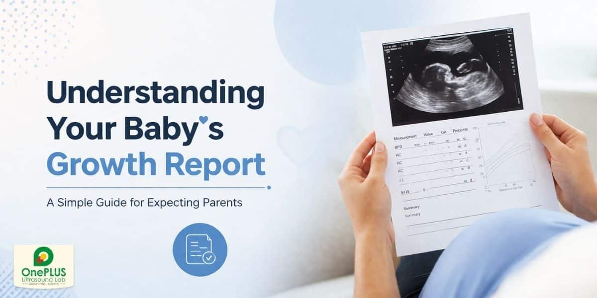
Key Measurements You’ll See in Your Report
When you open a growth scan report, the first section usually lists several measurements called fetal biometry measurements, the numerical backbone of your scan. These help doctors assess size and growth trends.
The most common measurements include:
- BPD (Biparietal Diameter) – the width of your baby’s head
- HC (Head Circumference) – the distance around the head
- AC (Abdominal Circumference) – the distance around your baby’s tummy
- FL (Femur Length) – the length of the thigh bone
Together, these create a picture of how proportionate and healthy your baby appears for your stage of pregnancy.
These numbers are often plotted against standard fetal growth charts to give a percentile, the value that helps show whether your baby’s size is typical, small, or large compared to other babies at the same growth scan weeks.
What Does “Normal” Look Like on a Growth Scan Report?
A growth scan normal report highlights measurements that fall within the expected range for your baby’s gestational age. In general:
- A percentile between 10th and 90th is usually considered within normal limits
- Percentiles below the 10th might suggest small size
- Percentiles above the 90th might suggest larger size
Keep in mind that “normal” isn’t the same for every pregnancy. Genetics, parental size, and health conditions like gestational diabetes or high blood pressure all influence how your baby grows. If a single measurement seems outside the normal range, it doesn’t always mean something is wrong, trends over time matter most.
Providers may recommend repeat scans to watch the growth curve rather than reacting to one data point.
Also Read: How to Read a Pregnancy Scan Report: A Guide For Patients
What Is Included in Third Trimester Growth Ultrasound Scan
A third trimester ultrasound tends to include more than just biometry. If your care provider ordered a growth scan, you’ll likely see:
- Estimated Fetal Weight (EFW) — calculated using formulas based on multiple measurements
- Amniotic Fluid Index (AFI) — shows the amount of fluid around the baby
- Placental position (important for birth planning)
- Baby’s position — such as head down (cephalic) or breech
These help give your doctor a more comprehensive picture of your baby’s readiness for birth and overall wellbeing. Each top parameter contributes to what doctors call a third trimester ultrasound report analysis.
How to Understand Fetal Growth Scan Report Step by Step
Let’s walk through how to read the main parts of your report:
Step 1: Check the Gestational Age
The report should confirm how many weeks along you are (e.g., 30 weeks + 2 days). This age is important because all size comparisons are based on it.
Step 2: Read the Fetal Biometry Section
Look for BPD, HC, AC, and FL. These measurements are compared with a standard to generate percentiles. Percentiles show where your baby stands compared to others at the same stage.
Step 3: Review the Estimated Fetal Weight
This shows your baby’s approximate weight. An anomaly scan baby weight estimate below or above the average might prompt closer monitoring, especially if it’s consistently low or high.
Step 4: Amniotic Fluid and Position
Normal fluid levels and a head‑down position are reassuring signs as you approach delivery.
Step 5: Placenta and Overall Notes
A note on the placenta’s location and any comments from the sonographer or doctor helps give context to all other measurements.
You can use this structured approach to go from “what is all this?” to “okay, I see where my baby stands!”
What If Something Seems Abnormal?
Don’t panic if a number is slightly outside a typical range. Ultrasound weight estimates can have a margin of error, sometimes up to 10‑15%, because babies don’t always stay still for perfect measurements.
Healthcare providers focus on patterns rather than single numbers. If a baby’s growth estimations seem low or high over consecutive scans, your provider might order additional monitoring or tests to explore potential causes.
In many cases, irregular values simply result from natural variation, and most babies land safely within a healthy range at birth.
Why Professional Interpretation Matters
Even though you can learn how to read a growth scan report, professional interpretation is vital. Experienced sonographers and doctors consider your full medical history, health conditions, and multiple factors when explaining what your results mean.
If you’re in the North Delhi area, trusted services like Ultrasound in Shalimar Bagh, and our earlier-mentioned Ultrasound in Rohini at OnePLUS Ultrasound Lab can help you understand your scans better. Visiting a centre with strong expertise means clarity and reassurance, not confusion.
Wrapping Up: How to Confidently Read Your Fetal Growth Scan Report
Interpreting a growth scan report doesn’t have to leave you puzzled. By learning how the key measurements work, what a “normal” scan shows, and how experts evaluate your baby’s growth, you can approach your pregnancy journey with confidence.
Remember:
- Growth scan weeks help compare your baby to standard growth charts.
- Fetal biometry numbers and percentiles show size and growth patterns.
- Estimated fetal weight gives you a ballpark of how much your baby might weigh near delivery.
- Follow‑up scans and professional interpretation are the best way to understand what the report means for your pregnancy.
Growth scans are tools, not verdicts, and most results end up reassuring new parents. Approach yours with curiosity, not fear!
Ready to get a clear, expert interpretation of your baby’s growth scan? Book your appointment today at OnePLUS Ultrasound Lab and gain peace of mind with trusted professionals guiding you every step of the way.
FAQ’s For Growth Scan Report
A standard anatomy ultrasound focuses on organ development and early pregnancy structure. A growth scan zeroes in on growth trends and size as the baby gets closer to birth.
Yes. Scans estimate weight using formulas that are useful but not perfect. A small margin of error doesn’t automatically mean concern.
For most low‑risk pregnancies, one scan in the third trimester is typical. If your doctor wants to track growth more closely, or if you have health risks, you may see more frequent scans.